
From Pixels to Life: Decoding Cellular Worlds with Cryogenic Electron Microscope – 19 March @ 6PM
IEEE CENTRAL COAST FREE EVENT – 19 March @ 6PM @ RUSTY’S PIZZA
“From Pixels to Life: Decoding Cellular Worlds with Cryogenic Electron Microscope”
Niels Volkmann, Prof., ECE, UCSB
Location – Rusty’s Pizza 5934 Calle Real, Goleta, CA 93117
5:00 PM – Complimentary Pizza, Salad, Beverage
6:25 PM – Central Coast Section Status
6:30 PM – Neils Volkmann Presents
Please join us March19th for an IEEE Central Coast Event when Prof. Niels Volkmann (UCSB) will present his lecture from the UCSB DISTINGUISHED LECTURE at the ECE SEMINAR SERIES
Link to Register yourself and Guests https://events.vtools.ieee.org/event/register/473688
“From Pixels to Life: Decoding Cellular Worlds with Cryogenic Electron Microscope”
Abstract: Cryogenic electron microscopy (cryo-EM) is a groundbreaking imaging technique that unveils cellular landscapes at the resolution of their molecular building blocks. This talk introduces the core principles of cryo-EM and highlights the vital role of signal processing and computational methods in transforming raw data into a deeper understanding of the machinery that drives life.
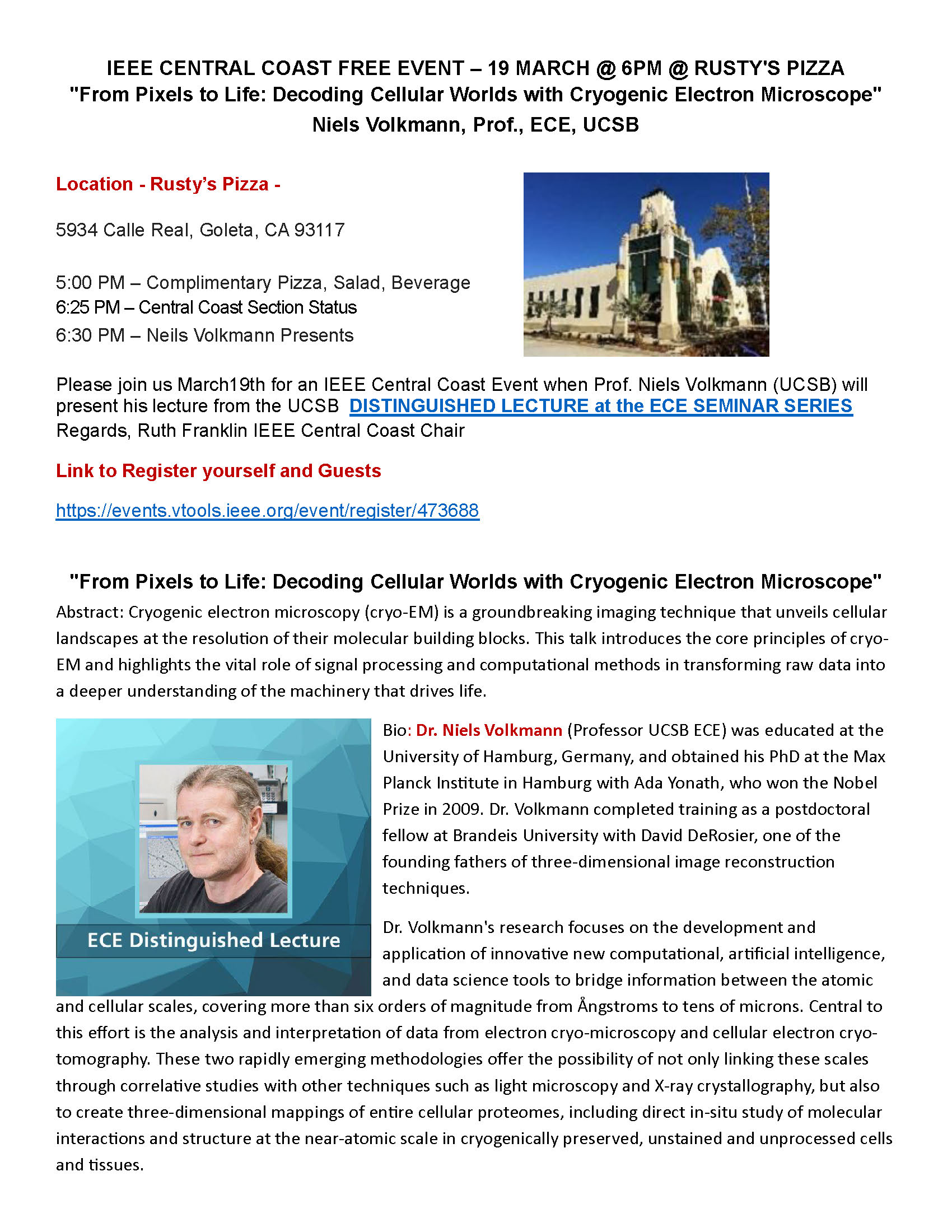
Bio: Dr. Niels Volkmann (Professor UCSB ECE) was educated at the University of Hamburg, Germany, and obtained his PhD at the Max Planck Institute in Hamburg with Ada Yonath, who won the Nobel Prize in 2009. Dr. Volkmann completed training as a postdoctoral fellow at Brandeis University with David DeRosier, one of the founding fathers of three-dimensional image reconstruction techniques.
Dr. Volkmann’s research focuses on the development and application of innovative new computational, artificial intelligence, and data science tools to bridge information between the atomic and cellular scales, covering more than six orders of magnitude from Ångstroms to tens of microns. Central to this effort is the analysis and interpretation of data from electron cryo-microscopy and cellular electron cryo-tomography. These two rapidly emerging methodologies offer the possibility of not only linking these scales through correlative studies with other techniques such as light microscopy and X-ray crystallography, but also to create three-dimensional mappings of entire cellular proteomes, including direct in-situ study of molecular interactions and structure at the near-atomic scale in cryogenically preserved, unstained and unprocessed cells and tissues.